| ■ |
Implants |
| |
| |
Material |
Characteristics |
Porosity |
Fabrication
method |
| CT-Bone |
α-tricalcium phosphate
(α-TCP) |
Un-sintered, connecting pores |
48% |
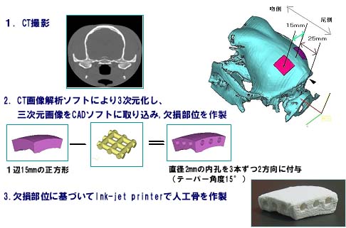
Three dimensional lamination method by ink-jet printing technology. |
| Existing artificial bone products |
Hydroxyapatite
(HA) |
Sintered porous material |
55% |
Cut by hand from the block |
|
| |
|
| ■ |
Fabrication method |
| |
 |
|
▲PAGE
TOP |
|
|
| ■ |
CT
findings |
|
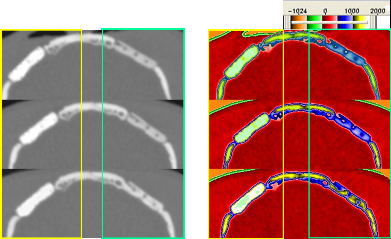
Narrowing of connecting macropores and bridge formation between CT-Bone and bone stump are observed. |
|
|
Immediately
after surgery
4 weeks after
24 weeks after
|
 |
|
Apacerum
CT-Bone
Apacerum
CT-Bone |
|
|
| ■ |
HU
analysis |
|
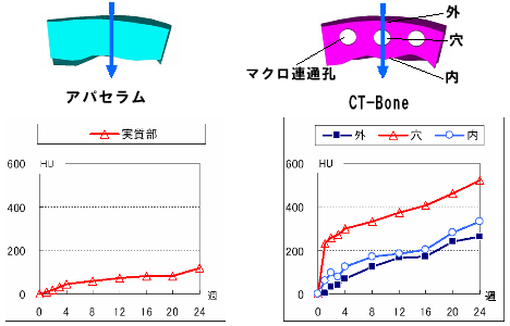
CT-Bone shows significantly more increment in HU than Apacerum. |
|
Profile
Line
Profile Line |
|
 |
|
▲PAGE
TOP |
| ■ |
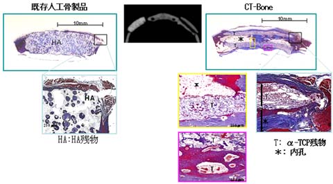
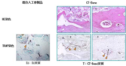
Histologic findings |
|
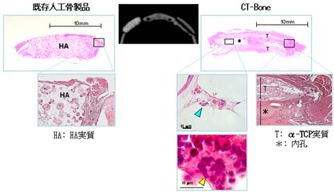
(1)HE
staining
In Apacerum, osteocytes were found only in its body.
In CT-Bone, bone-like tissue penetrates into connecting macropores. In addition, bone marrow formation was observed, with erythroblasts pointed by the blue arrow and megakaryocytes pointed by the yellow arrow. |
|
 |
|
(2)MT
staining
Blue collagenous fibers and bone tissues penetrate into Apacerum body and mactopores of CT-Bone. |
|
 |
|
(3)TRAP
staining
TRAP stain shows osteoclasts pointed by the orange arrow. |
|
 |
|
▲PAGE
TOP |
| ■ |
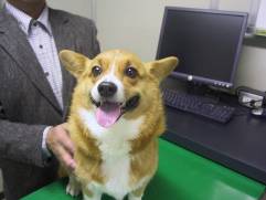
Application to animal patient |
|
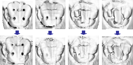
・Corgi Dog suffering from bone sarcoma underwent tumor resection, and CT-Bone was implanted to the bone defect site.
・Granulomas was removed in implantation.
Implant was performed on 19th July, 2005.
|
|
・The patient dog has been followed up by blood tests and CT scanning, showing steady recovery.
|
|
 |
|
The implanted CT-Bone
The patient dog looks fine. |
|
|
|
・Temporal profile of CT of the implanted site |
| |
Immediately
after surgery 3 months after
7 months after 10
months after |
| |
|
HU339〜3071
HU110〜3071 |
 |
| |
|
| |
▲PAGE
TOP
|